Examination of the patient, is the main thing to determine correct diagnosis necessary for successful treatment in the future. Before patient treatment stomatologist should find out about the patient required information for correct planning and carrying out of treatment. The examination methods used in our clinic by team of specialists are the best nowadays.
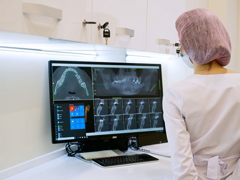
Radiography of pathology of the teeth, jaws, other bones of the face, maxillary, frontal sinuses, temporomandibular joints, sinuses of the mouth are important, because they are the main diagnostic method of disease. In our clinic, we perform intraoral and extraoral X-ray images. According to X-rays images, it is possible to judge condition of bone tissue surrounding tooth and bones of maxillofacial area. In case of treatment of several teeth X-ray load risk is fully minimized due to panoramic radiography – method, by which it is possible to get image of all the teeth and bone tissue on a film. One more method of by three-dimensional study of the oral cavity, bone and soft tissue is a modern computer tomography (CT) scan, which uses X-rays. The ray passes through the patient's body, getting on a special detector. Getting such three-dimensional image doctor with high accuracy can identify position, shape, size and structure of the various structures of the body. Because of this, CT is one of the key instruments of modern diagnostics in stomatology.
Obligatory method of inspection in orthodontics is using of cephalometric analysis for diagnostic and planning of the treatment. Cephalometry helps our doctors orthodontists, orthopedists, maxillofacial surgeon, implant surgeon and physicians to understand better expectations of the patient. For example, knowing restrictions and possibilities of orthodontic treatment it is possible to assign clearly tasks to orthodontist during the preparation of the patient for implantation and subsequent prosthesis. Cephalometric analysis expands boundaries of the understanding of stomatological problems and thanks to that; specialists of our clinic are able to “see” the problem on not only the level of teeth and teeth arches, and the patient face as a whole. This allows us to understand what changes we should expect during the treatment.
Teleroentgenography is method of radiological research, which uses for study of the structure of the facial skeleton, specification of the diagnosis and prognosis of orthodontic treatment, to detect changes that arise during the treatment. In direct projection, TEG gives an opportunity to diagnose abnormalities of maxillodental system. On the TEG image bones of the facial skull and soft tissues borders are displayed, that allows to study their correlation. Except orthodontics, teleroentgenography successfully uses in surgery to diagnose fractures and cracks. With help of the images doctor can determine exactly asymmetry of the facial bones, variety of the inflammatory diseases, as well as the fractures and displacement of the nasal septum. TEG is an important part of stomatological examination, in which doctor can make diagnosis and plan treatment.
Modern stomatology uses digital photography. Typically, standard kit includes diagnostic pictures of intermediate and final stages of the treatment. Diagnostic pictures are the key things. It is possible to define shape of future teeth, their color, there is a choice of optimal construction in terms of aesthetics, and is facilitated interaction in the chain of patient-doctor-technician. Intermediate images has scientific and statistic content. It depends of where they are – in the doctor’s computer or in his article in the magazine. It is worth to mention that in some cases after thorough study these images give an opportunity to see all shortcomings in the work, and will prompt how to solve them. Final images are extremely important as provide an opportunity to evaluate the result.
Diagnostics in the articulator uses in primary examination of the patient and on the stages of treatment (during prosthesis, restoration, orthodontic treatment).
Articulator is a tool reflecting movements of mandible. This analysis allows doctor to analyze moves in temporomandibular joint, displacement of the articular bone heads, consider occlusal contacts, position of the mandible in habitual occlusion and in the central correlation. With help of articular diagnostic, our doctors will determine usual occlusion of the patient; will install his occlusion problems that will allow determining start position for future treatment. Using articulator gives opportunity to trace movements of mandibular relative to maxilla and study centric eccentric occlusion.
For accurate elevation of equilibrium function of the body, our doctors perform postural (osteopathic) tests, which are extremely important for patients with dentition anomaly. Purpose of the study of body of the patient by the orthodontic is objectification of progress changes after orthodontic treatment.
Kinesiology (muscle) test is one of the main instruments of homeopathy-synergy medicine that allows with help of tonus muscle study evaluate of the vitality of the body or its systems. Due to this test patient starts to understand himself and appears a choice: leave everything as it was or change life for the best. Every person is free to choose his life path if this choice is deliberate.